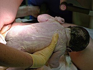
Vernix caseosa, also known as vernix, is the waxy white substance found coating the skin of newborn human babies.[1] It is produced by dedicated cells and is thought to have some protective roles during fetal development and for a few hours after birth.
Etymology
In Latin, vernix means varnish and caseosa means cheesy. The term was first published in 1846 in the Dunglison Dictionary of Medical Sciences.[1]
In-utero development
Vernix is produced during a distinct phase of the epidermal development.[2] Around the 21st week of gestation, periderm cells are being shed and replaced with strateum corneum; these shedding mix with secretions of sebum by the sebaceous glands to form vernix, which gradually covers the body in an anteroposterior and dorsoventral pattern.[1][2][3] Vernix, in itself, is also believed to aid in the formation of strateum corneum.[4] By early third trimester, the process is complete.[5]
Soon enough, part of the vernix is emulsified by increasing concentrations of pulmonary surfactants and desiccates, only to be consumed by the fetus; a corresponding increase in amniotic fluid turbidity is noticed.[2]
Characteristics
Composition
Vernix has a highly variable makeup but is primarily composed of sebum, cells that have sloughed off the fetus's skin and shed lanugo hair.[6] Chemically, it is water (80%), lipids (10%) and proteins (10%).[1] The lipids include ceramides, cholesterol, fatty acids, triglycerides, waxes and sterol esters, squalene, and phospholipids;[1] multiple detailed analyses of the polar components have been done.[7] The protein composition is relatively understudied.[1] Vernix of term infants has more squalene and a higher wax ester to sterol ester ratio than preterm infants.[6]
Morphology
Vernix is composed of mobile corneocytes embedded in an amorphous lipid matrix.[1] Precise biological mechanisms leading to its formation are hazily known.[8]
The cells are polygonal or ovoid in shape, malleable, and lack nuclei; typical thickness is 1-2 µm.[1] Nuclear ghosts are frequently observed and Acid Phosphatase Activity is nonuniform.[1] Keratin filaments build a scaffold like structure which form a water-storage area.[1] As opposed to stratum corneum, the vernix corneocytes lack desmosomal attachment and the lipid layer is more disordered.[9]
Physical properties
Vernix is a white viscous cream-like substance in appearance.[1]
The water is not uniformly distributed throughout, but rather exclusively present in the sponge-like corneocytes; despite its high water content, vernix is non-polar (due to lipids) and more vapor-permeable than strateum corneum.[1][10][11]
Functions
Vernix appears in all full term infants but with widely varying body-coverage, while premature and post-mature births generally do not display any.[6][2][12]
It is theorized (and observed) to serve several purposes:[1][2][10]
- Electrical isolation of the fetus (this affects accurate fECG measurement of fetal heartbeat).[13]
- Waterproofing the skin, whilst in gestation.
- Lubricating the infant's skin, and facilitating easy passage through the birth canal.
- Preventing infections — primarily as a mechanical barrier and secondarily via the presence of lysozyme, lactoferrin and antimicrobial components in peptide layer.
- Moisturizing the stratum corneum whilst in gestation (and controlled drying in post-partum phase).
- Thermoregulation in post-partum phase — evidence is mixed.
- Quick healing of epidermal wounds.
- Development of gut, after intra-uterine consumption.
Medical uses
Vernix is used as a reliable site-of-record for measuring cocaine exposure in pregnant women.[2] Using vernix for diagnosing uterine rupture and amniotic fluid embolism has been proposed.[2]
Disorders
Granuloma and peritonitis of vernix have been observed in Caesarean sections.[2] High volumes of vernix cause Neonatal Aspiration Syndrome.[2]
Other species
Vernix is thought to be unique to human fetal development; in 2018, vernix-like material was reportedly obtained from pups of Zalophus californianus.[14]
Additional images


This article uses material from the Wikipedia article
Metasyntactic variable, which is released under the
Creative Commons
Attribution-ShareAlike 3.0 Unported License.
Metasyntactic variable, which is released under the
Creative Commons
Attribution-ShareAlike 3.0 Unported License.