Lck (or lymphocyte-specific protein tyrosine kinase) is a 56 kDa protein that is found inside specialized cells of the immune system called lymphocytes. Lck is a tyrosine kinase, which phosphorylates tyrosine residues of certain proteins involved in the intracellular signaling pathways of these lymphocytes. It is a member of the Src family of tyrosine kinases.


T cell signaling
Lck is most commonly found in T cells. It associates with the cytoplasmic tails of the CD4 and CD8 co-receptors on T helper cells and cytotoxic T cells,[5][6] respectively, to assist signaling from the T cell receptor (TCR) complex. When the T cell receptor is engaged by the specific antigen presented by MHC, Lck acts to phosphorylate the intracellular chains of the CD3 and ζ-chains of the TCR complex, allowing another cytoplasmic tyrosine kinase called ZAP-70 to bind to them. Lck then phosphorylates and activates ZAP-70, which in turn phosphorylates another molecule in the signaling cascade called LAT (short for Linker of Activated T cells), a transmembrane protein that serves as a docking site for a number of other proteins, the most important of which are Shc-Grb2-SOS, PI3K, and phospholipase C (PLC). Additionally, upon T cell activation, a fraction of kinase active Lck, translocates from outside of lipid rafts (LR) to inside lipid rafts where it interacts with and activates LR-resident Fyn, which is involved in further downstream signaling activation.[7][8]
The tyrosine phosphorylation cascade initiated by Lck and Fyn culminates in the intracellular mobilization of calcium (Ca2+) ions and activation of important signaling cascades within the lymphocyte. These include the Ras-MEK-ERK pathway, which goes on to activate certain transcription factors such as NFAT, NF-κB, and AP-1. These transcription factors regulate the production of a plethora of gene products, most notable, cytokines such as Interleukin-2 that promote long-term proliferation and differentiation of the activated lymphocytes. In addition to significance of Lck and Fyn in T cell receptor signaling, these two src kinases have also been shown to be important in TLR-mediated signaling in T cells.[9]
The function of Lck has been studied using several biochemical methods, including gene knockout (knock-out mice), Jurkat cells deficient in Lck (JCaM1.6), and siRNA-mediated RNA interference.
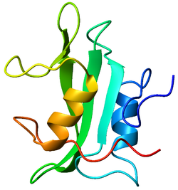
Structure
Lck is a 56-kilodalton protein. The N-terminal tail of Lck is myristoylated and palmitoylated, which tethers the protein to the plasma membrane of the cell. The protein furthermore contains a SH3 domain, a SH2 domain and in the C-terminal part the tyrosine kinase domain. The two main phosphorylation sites on Lck are tyrosines 394 and 505. The former is an autophosphorylation site and is linked to activation of the protein. The latter is phosphorylated by Csk, which inhibits Lck because the protein folds up and binds its own SH2 domain. Lck thus serves as an instructive example that protein phosphorylation may result in both activation and inhibition.
Substrates
Lck tyrosine phosphorylates a number of proteins, the most important of which are the CD3 receptor, CEACAM1, ZAP-70, SLP-76, the IL-2 receptor, Protein kinase C, ITK, PLC, SHC, RasGAP, Cbl, Vav1, and PI3K.
Inhibition
In resting T cells, Lck is constitutively inhibited by Csk phosphorylation on tyrosine 505. Lck is also inhibited by SHP-1 dephosphorylation on tyrosine 394. Lck can also be inhibited by Cbl ubiquitin ligase, which is part of the ubiquitin-mediated pathway.[10]
Saractinib, a specific inhibitor of LCK impairs maintenance of human T-ALL cells in vitro as well as in vivo by targeting this tyrosine kinase in cells displaying high level of lipid rafts.[11]
Masitinib also inhibits Lck, which may have some impact on its therapeutic effects in canine mastocytoma.[12]
| This article uses material from the Wikipedia article Metasyntactic variable, which is released under the Creative Commons Attribution-ShareAlike 3.0 Unported License. |